
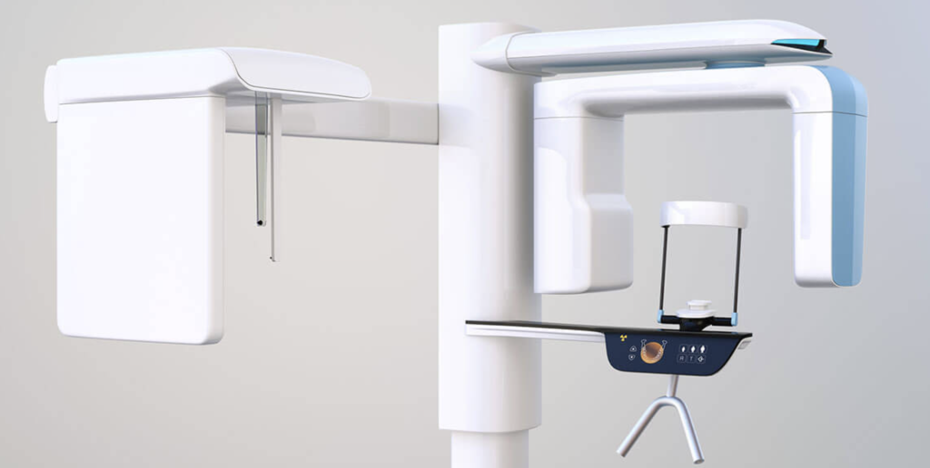
What is Cone Beam Computed Tomography?
CBCT uses a “cone-shaped” X-ray, instead of a “fan-shaped” beam. This allows oral and maxillofacial structures to be captured in two dimensions. With a cone-shaped beam, diagnostic information can be found faster and at a lower radiation dose. This way, more complex diagnostic and treatment planning can be completed in a single scan.
Why is Cone Beam CT different from Traditional X-Rays?
Cone Beam CT is easier and more cost-effective than a conventional “CT” scan or X-RAY. It allows your dentist to produce high-quality three dimensional images of the teeth and jaw. Compared to CT scans, cone beam imaging provides better visualization, more accurate scans, and lower levels of radiation. It’s an increasingly popular choice compared to traditional X-rays, as it can create a better process for the patient and surgeon.
Dental Care Using CBCT
CBCT scanners are also more accessible to dental offices. They are less expensive, smaller, lighter, and offer better spatial resolution overall. Having a 3D view allows dentists to see anatomical structures, produce high-quality images from different angles, and create detailed surgical treatment plans.
Here are just some of the potential dental applications for CBCT three-dimensional imaging:
- Assessment of bone volume for implant placement
- Preparing for surgery to address impacted teeth
- Planning for more than one complex implant surgery virtually
- Discovering airway analysis for sleep apnea
- Planning for bone reconstruction or grafting
- Measuring proximity to important structures like the sinus and nerve pathways
- Measuring the size of growths, tumors, or cysts in the jaws.
- Planning jaw surgery virtually
- Diagnosing TMJ
- Locating impacted teeth in relationship to the skeleton
- Diagnosing facial fractures and performing facial reconstruction.
Cone beam CT provides clearer, more detailed images than regular dental X-rays, and can improve care across a wide range of procedures.
Do I need to prepare for a CBCT scan?
Heavy preparation shouldn’t be necessary for cone-beam computed tomography. You may need to remove items that will interfere with imaging, such as jewelry, hearing aids, or removable dental work. It’s also important to inform your surgeon if there is a possibility that you may be pregnant. Some scans are taken with the patient lying down, others are taken seated. A CBCT scan only takes 5 to 40 seconds. All patients need to do during the procedure is bite down in a specific jaw position, and then sit as still as possible. During the procedure, the scanner will rotate around the patient’s head to take images that will be combined into a 3-D image. During the scan, 150 to 200 images will be taken. The procedure is completely painless and noninvasive. After the procedure, patients can go about their day as normal. Your oral surgeon will analyze the images and contact you with the results and next steps.
Nervous about CBCT Radiation Levels?
Are you concerned about radiation exposure with CBCT imaging? Cone beam CT actually provides significantly less radiation exposure than traditional dental X-rays. CBCT can be a very low-dose modality compared to traditional medical imaging. Dosages are also progressively decreasing. CBCT technology allows for an accurate diagnosis with a relatively low dose of radiation. An upper or lower jaw scan to prepare for and diagnose dental issues is about 95 µSv. Comparatively, a coast-to-coast round-trip flight is 30 µSv, so if you travel frequently, this isn’t a big difference! Traditional CT scanners are much higher in radiation. A regular mammogram is over 4 times the µSv of a CBCT jaw scan. A CT of your abdomen or pelvis clocks in at 10,000 µSv. It’s understandable to be concerned about radiation with CT scans, but remember that this one is relatively minor. Remember that your dentist is as concerned with your health as you are.
3D Cone Beam Imaging at Wilshire Oral Surgery
If you’re looking for state-of-the-art technology and a smooth dental experience in Los Angeles, check out Wilshire Oral Surgery. Wilshire’s oral surgeons truly care about patient outcomes and are committed to providing quality care. CBCT imaging technology is commonly used at Wilshire, and our surgeons are committed to providing a positive and safe experience. Choose a dental office that provides more with Wilshire.